When a dog suddenly yelps during exercise or displays acute hindlimb lameness—holding the affected leg elevated off the ground—it rarely stems from a simple sprain. It often points to a critical orthopedic condition known as canine CCL rupture (cranial cruciate ligament rupture). Unlike human cruciate tears that typically result from acute traumatic hyperextension, canine CCL disease is predominantly an insidious, chronic degenerative condition driven by abnormal biomechanical loading. Left untreated, cranial tibial subluxation causes secondary meniscal tears and irreversible osteoarthritis. This clinical report details the biomechanical pathogenesis, orthopedic diagnostic modalities, TPLO versus conservative management algorithms, and post-injury rehabilitation regimens.
1. Biomechanical Function and Mechanism of CCL Rupture
The canine stifle (knee) joint diverges fundamentally from human anatomy. While the human tibia sits nearly perpendicular to the femur during standing, the canine stifle remains in constant flexion, positioning the tibial plateau at a natural posterior-downward slope of approximately 20° to 30°.
Tibial Plateau Angle (TPA) and Cranial Tibial Thrust
Whenever a dog bears weight on a hindlimb, axial force drives the femoral condyles to slide down the sloped tibial plateau. The internal anatomical constraint counteracting this shearing vector is the Cranial Cruciate Ligament (CCL).
- Continuous running, accelerating, and jumping generate persistent tensile stress along the ligament’s collagen bundles.
- Steeper Tibial Plateau Angles (TPA) directly amplify the cranial tibial thrust force.
- In large breeds and overweight dogs, this constant load causes progressive microscopic fatigue of the ligament fibers, eventually culminating in rupture under routine, non-traumatic movement.
High-Risk Cohorts and Predisposing Factors
- Genotypic and Conformational Risk: Working and large breeds including German Shepherds, Labrador Retrievers, Rottweilers, and large shepherd mixes exhibit elevated prevalence due to body mass and high energy output.
- Contralateral Rupture Incidence: Approximately 40% to 50% of dogs experiencing unilateral CCL rupture suffer a contralateral rupture within 12 to 24 months as a consequence of compensatory weight shifting.
- Obesity and Sarcopenia: Deficits in stabilizing musculature (quadriceps, hamstrings) paired with elevated body fat multiply static mechanical strain on the passive ligamentous stabilizers.
2. Clinical Differentiation and Orthopedic Diagnostic Criteria
Canine cruciate disease presents across a continuum from partial tears to complete ruptures accompanied by meniscal displacement.
| Clinical Parameter | Partial Tear (Early) | Complete Acute Rupture | Secondary Meniscal Injury |
| Gait Evaluation | Post-rest stiffness, exercise lameness | Non-weight-bearing hindlimb lameness | Severe toe-touching, audible joint click |
| Sitting Posture (Sit Test) | Minor outward abduction of limb | Inability to flex stifle; leg extended laterally | Refusal to sit; persistent standing |
| Palpatory Stifles | Medial buttress (capsular thickening) | Marked cranial tibial subluxation | Severe pain upon full flexion/extension |
Diagnostic Workup in Veterinary Practice
- Cranial Drawer Test: Manual immobilization of the distal femur while applying cranial translation to the proximal tibia. Objective sliding indicates loss of CCL functional integrity.
- Tibial Compression Test: Flexion of the hock joint mimics axial weight-bearing; forward displacement of the tibial tuberosity confirms pathological cranial thrust.
- Orthopedic Radiography: Rules out neoplasia or avulsion fractures, identifies infrapatellar fat pad displacement, quantifies joint effusion, and precisely measures the Tibial Plateau Angle (TPA) for surgical planning.
3. TPLO Osteotomy vs. Conservative Medical Management
Because a torn canine cruciate ligament does not self-repair, treatment pathways depend strictly on patient weight, stifle geometry, and lifestyle demands.
Mechanistic Principles and Indications for TPLO
In medium-to-large canines (>15 kg), static suture techniques (Extracapsular Lateral Suture) exhibit high failure rates due to massive tensile loads. Consequently, Tibial Plateau Leveling Osteotomy (TPLO) stands as the international gold standard.
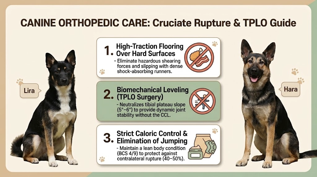
- Surgical Mechanism: A radial cut is performed on the proximal tibia, the plateau is rotated to level the slope to approximately 5°–6°, and the segment is rigidified using a dedicated internal bone plate.
- Clinical Superiority: By neutralizing the sloped anatomy, gravitational load itself stabilizes the joint dynamically during stance phase. The dog achieves functional stability without needing an intact ligament.
Limitations and Criteria for Conservative Management
- Reserved strictly for patients with severe systemic comorbidities precluding anesthesia, or small-breed canines (<7 kg) with stable partial tears.
- Requires 8 to 12 weeks of rigid crate confinement, custom orthotic stifle bracing, multi-modal analgesics (NSAIDs, gabapentin), and disease-modifying osteoarthritis drugs (polysulfated glycosaminoglycans).
- In large breeds, conservative management frequently yields accelerated osteoarthritis, chronic pain, and over a 70% rate of secondary meniscal cartilage fragmentation.
4. Postoperative Environmental Management and Relapse Prevention
Long-term joint prognosis depends on post-surgical environmental control during the 8-to-12-week osteotomy healing window.
- High-Traction Indoor Flooring: Cover tile, hardwood, and vinyl floors with non-slip interlocking rubber runners (minimum 5 mm thickness) to prevent abduction slips and implant strain.
- Rigorous Caloric and Weight Control: A 5% increase in body weight accelerates wear on the contralateral limb. Calibrate diet portions to maintain a lean Body Condition Score (BCS 4/9).
- Restricted Leash Walking: Eliminate unconstrained running, jumping, and stairs for the first 6 to 8 weeks. Keep elimination walks controlled on a short 1.5-meter leash.
- Chondroprotective Supplementation: Administer pharmaceutical-grade Omega-3 fatty acids (high EPA/DHA concentrations) alongside glucosamine and chondroitin to suppress synovitis and protect cartilage.
Frequently Asked Questions (FAQ)
Q1. My dog limped for two days, rested, and is now walking normally. Is veterinary evaluation necessary?
Yes. In partial tears, periarticular inflammation temporarily wanes with rest, masking instability. However, uncorrected joint shear continues to destroy the articular cartilage and menisci. Any transient hindlimb lameness warrants immediate diagnostic palpation.
Q2. Do TPLO bone plates and screws require surgical removal later in life?
TPLO implants are made of biocompatible medical-grade titanium or surgical stainless steel and are designed to stay permanently in the bone. Surgical retrieval is indicated only in rare cases of delayed infection, cold sensitivity, or implant rejection.
Q3. How high is the risk of the opposite leg tearing after surgery?
Approximately 40% to 50% of large-breed dogs tear the contralateral cruciate ligament within one to two years. The uninjured limb bears unequal compensatory weight during the initial convalescence. Weight control and slip prevention remain the primary defense against bilateral breakdown.
Meta Description: Canine Cranial Cruciate Ligament Rupture (CCLR) guide: clinical lameness signs, sitting test indicators, TPLO surgery mechanics vs conservative care, and recovery protocols.