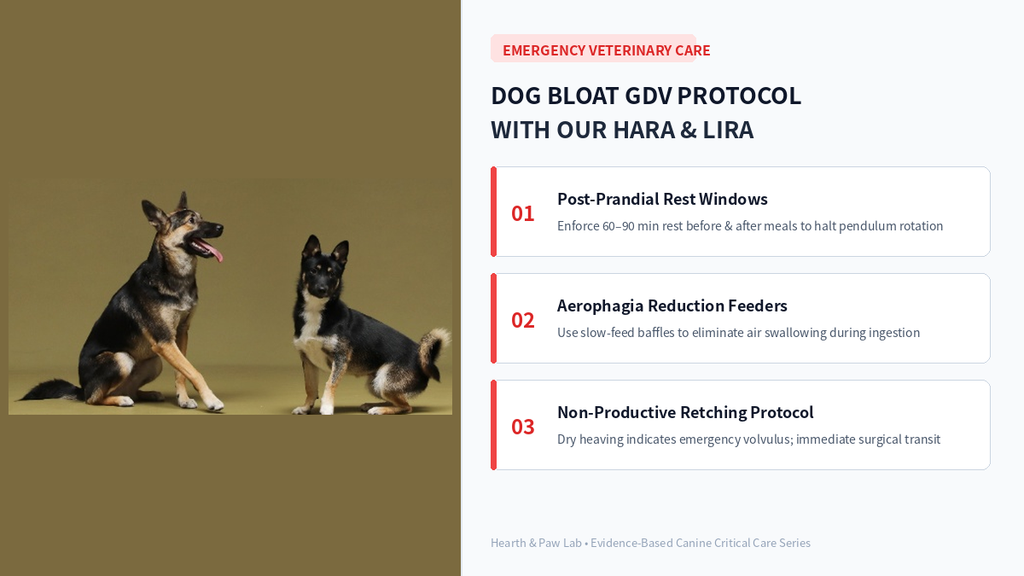
When executing disciplined dog bloat prevention, observing a canine repeatedly heave without producing vomitus alongside rapid abdominal distension signals Gastric Dilatation-Volvulus (GDV)—the most lethal gastrointestinal emergency in veterinary medicine. Deep-chested large canines and working mixes possess prolonged gastric ligaments, rendering their anatomically heavy stomachs susceptible to rotating 180 to 360 degrees along the mesenteric axis. This catastrophic twisting obstructs the caudal vena cava, sending the animal into hypovolemic collapse and rapid myocardial ischemia.
1. Pathophysiology of Canine Gastric Dilatation-Volvulus (GDV)
Aerophagia, Distension, and Rotation
Canines with vigorous appetites rapidly ingest dry kibble alongside copious air—a state termed aerophagia. The heavy, fluid-filled, and fermenting gastric chamber acts as a pendulum. A single torsional motion causes the pylorus to shift ventral to the fundus, completely occluding both the lower esophageal sphincter and the duodenal exit. Trapped intraluminal gas rapidly expands, severely increasing intramural gastric pressure.
Systemic Vascular and Splenic Sequelae
As intragastric pressure escalates, it compresses the portal vein and caudal vena cava, severely diminishing venous return to the heart. Cardiac output plummets, precipitating refractory distributive shock. Concurrently, the short gastric arteries to the spleen tear or twist, causing severe splenic congestion and triggering ventricular arrhythmias—the leading cause of post-decompression mortality.
Veterinary Insight: Non-productive retching—intense abdominal contractions yielding only white mucosal froth without vomitus—remains the hallmark indicator of true volvulus. This presentation mandates immediate emergency decompression and intensive surgical stabilization.
2. Comparative Matrix: GDV Risk Factors vs. Clinical Interventions
| Management Category | High-Risk Traditional Routine | Evidence-Based Preventive Standard | Expected Physiological Outcome |
| Feeding Frequency | Single massive daily meal | 2 to 3 metered rations daily | Avoids critical gastric volume overload |
| Ingestion Velocity | Unrestricted bowl ingestion | Maze baffles (Slow-Feeders) | Diminishes swallowed aerophagia by >70% |
| Post-Prandial Phase | Vigorous running/play post-meal | 60–90 minute enforced quiet window | Eliminates rotational pendular torque |
| Water Accessibility | Unrestricted ice water post-kibble | Metered lukewarm hydration | Prevents acute dry food expansion and spasm |
3. Systematic 3-Phase Gastrointestinal Safety Protocol
Phase 1: Enforced 90-Minute Post-Prandial Rest Windows
A full stomach acts as an unstable pendulum. Allowing high-impact exercises—such as frisbee, rapid turns, or stairs—immediately after feeding mechanically initiates gastric torsion. Maintain strict confinement or crate rest for at least 60 to 90 minutes post-feeding. Conversely, withhold heavy meals for at least 45 minutes following rigorous physical training until panting ceases completely.
Phase 2: Elimination of Feeding Elevation and Diet Auditing
Historical literature advocated elevating food bowls for large breeds; however, broad epidemiological surveys revealed that excessive bowl elevation actually increases air gulping and multiplies GDV incidence. Keep feeding vessels at floor level. Furthermore, scrutinize nutritional profiles: avoid commercial kibbles where fat or animal grease constitutes the primary surface top-dressing, as this delays gastric emptying times.
Phase 3: Prophylactic Laparoscopic Gastropexy
For large breeds with high thoracic-depth-to-width ratios or a documented family history of GDV, prophylactic gastropexy offers life-saving protection. Often performed during elective sterilization, this surgical procedure permanently adheres the gastric antrum to the right abdominal wall. While it does not halt gas accumulation (dilatation), it permanently eliminates fatal organ rotation (volvulus), dropping mortality rates below 5%.
Frequently Asked Questions (FAQ)
Q1. How can I differentiate simple dietary indiscretion from acute GDV?
Canines suffering from mild gastritis readily discharge bile, saliva, or food matter and subsequently relax. In contrast, a canine undergoing GDV exhibits distress, paces incessantly, salivates heavily, and displays an unyielding, drum-like tautness in the cranial abdomen.
Q2. Does a slow-feed bowl completely eliminate the risk of bloat?
No single tool acts as a total cure. Slow feeders directly curtail aerophagia, but familial genetic predispositions, autonomic nervous system stress, and post-prandial exertion remain strong co-factors requiring unified management.
Effective dog bloat prevention requires structural mealtime discipline. By enforcing a strict 90-minute resting perimeter, splitting caloric intake across multiple servings, and utilizing slow-feed matrices, you insulate your dog against the devastating progression of acute gastric dilatation-volvulus.
- Meta Description: A clinical guide to dog bloat prevention: identifying acute GDV symptoms, non-productive retching, feeding protocols, and critical resting windows.